Episode Transcript
Announcer: Examining the latest research and telling you about the latest breakthroughs, The Science and Research Show is on The Scope.
Interviewer: Each year the National Institutes of Health gives a New Innovator award for high risk, high impact research. Dr. Adam Frost from biochemistry at the University of Utah received the award for coming up with innovative ways to visualize the tiny machines that do the essential work in our body's cells. Dr. Frost, how did it feel to get the award?
Dr. Adam Frost: Thrilling, but even more than that, relaxing. I felt like I could let my guard down or unwind a little bit for the first time in many years.
Interviewer: Why is that? There's a stress for funding?
Dr. Adam Frost: Yeah, there was certainly a great deal of stress starting my own lab, becoming an assistant professor, hiring people, and having the resources to both pay them and do great science. Now we've got that. We should be okay for about five years to do the research we want to do.
Interviewer: So explain your research.
Dr. Adam Frost: It's actually quite hard to study cell biology at a certain scale, at a certain dimension. A lot of beautiful work is done at the near atomic scale where we isolate a single molecule and look at it on the length scale of what are called angstroms, the average distance between, say, two carbon atoms in a molecule. But then, if you want to go to the next scale up where you, say, take one, two, or a hundred proteins and have them come together to build a machine in the cell that has a certain job, that gets tough.
It gets tough biochemically to isolate these elaborate machines. It's tough genetically to understand all their moving parts, and it's very tough structurally because these things can become so big and also so flexible that traditional structural approaches are basically impossible. Yet they're still a little bit too small to see with a light microscope. So I'm talking about things that are tens of nanometers by tens of nanometers. It's at that length scale that experiments are very challenging to do but where my lab is seeking to apply our particular expertise.
Interviewer: Are there applications for determining what these structures look like and what they do?
Dr. Adam Frost: The New Innovator grant was concerned with how cells divide. I'm particularly interested in this because it's this fundamental event in which a mother cell becomes two daughter cells and somehow everything that each daughter cell needs is inherited and the cells separate without any leaking or loss of integrity. So my lab took our now somewhat familiar approach of identifying the genes genetically, then purifying the components biochemically.
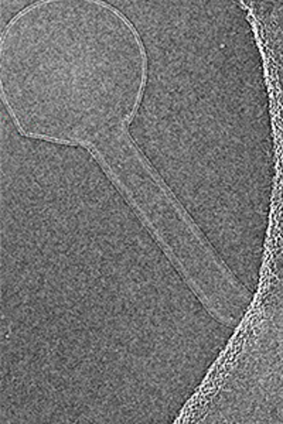
In this particular case I want to give a lot of credit to my department chair, Dr. Wesley Sunquist, and a postdoctoral fellow in his lab, John McCullough. Together, we were able to, in the test-tube, recreate the machinery that helps drive cell division. This was tough in that it's a complicated machine with many pieces and not only do they have to assemble with each other, they have to bind to a membrane and reshape the membrane into this thin tunnel that connects two daughter cells before ultimately twisting that tunnel together so that it can break with no leaking.
We've been able to successfully recreate this phenomenon in the test-tube, take micrographs of it in a modern electron microscope where we can actually see each of the proteins involved. So for many, many thousands, and in some cases more, images we use computers and computer algorithms to recreate a three-dimensional structure from lots and lots of two-dimensional pictures. It's a process that's a little bit analogous to having a CAT scan where your brain is imaged from lots of different angles and each of those images can be used in the computer to build up a three-dimensional structure of your brain, your heart, or whatever you are having imaged in a CAT scan. We do the same thing with molecules so that we can build up a three-dimensional structure of a molecule for many, many, many two-dimensional pictures.
Interviewer: What is the high risk aspect of this work?
Dr. Adam Frost: Most people stay away from these because they're hard to work with. In contrast to proteins that are perfectly happy to just live in the test-tube, membrane proteins and membrane binding proteins really are only happy when on or in a membrane. That adds a new dimension to the biochemical side of the project that most people would prefer to avoid.
One of the other things that's risky or challenging about this work is, as I alluded to before, it's expensive. While I was a postdoctoral fellow, for example, we learned that all of our molecules were moving while we were taking their pictures and that the real limit to our resolution was that over, say, a one-second exposure our molecules were dancing around and our final images were therefore quite blurry. So very recently ultrafast detectors have been invented. These ultrafast detectors actually allow us to de-blur the motions involved and get crystal-clear images of these molecules.
So it's risky in that the experiments are quite challenging. It's expensive in that these reagents or uncommon and many we have to make de novo, but I think we were able to convince the NIH that while it was risky and most people don't work on this problem we've made strong progress, both in the biochemistry and in the imaging and the image analysis problems and that we're well on our way.
Interviewer: What is the significance? What can you do with this information?
Dr. Adam Frost: I have a couple of answers to that question. My first answer is always to say that we don't always know the end from the beginning when there's a mystery. When we look at something like cell division and we can watch a movie of cells dividing or we can, just with our own eyes, look in the microscope and see cells go through this incredible process of division, and I see one of our jobs as to replace that sense of awe and miracle with a sense of understanding. How that understanding will take us to new places technologically is often very hard to predict.
A standard example of that is the people who invented the transistor had no idea it would lead to computers that we all now depend on constantly. So I have high hopes that at any time that we can take a fundamental mystery about biology especially and replace that mystery with a genuine understanding. That will have impacts in medicine that we probably can't predict from where we're sitting today.
But in the case of the cell division machinery I can actually be a little bit more concrete. Believe it or not, the machinery that drives cell division is found in other contexts as well. Again, my chair, Wes Sunquist, and several other labs showed that pathogens, especially viruses like HIV, highjack these same genes, the same machinery in their infectious process. So everything we're learning and that I've described about the mechanism of cell division actually applies to how HIV buds from and escapes from infected cells to go and infect additional cells. So this thin, membranous neck that cells pass through when they divide, viruses have to create the same neck as they escape from an infected cell. So, again, anything we can learn about the chemical mechanism, the atomic details, of how this machinery works will tell us how human pathogens infect and escape from human cells.
Interviewer: I'm struck by the fact that you actually have a medical background and your work is actually pretty far removed from the clinic, although obviously it does have some health applications. What perspective does your medical background give you that other basic researchers in this field do not have?
Dr. Adam Frost: Well, I'm not sure. I don't want to claim any special insight. I definitely feel that my MD shapes the way I think and gives me a certain background in human pathology that helps me weight problems differently. While as you say, my lab is pretty far removed from the clinic, we tend to only want to work on problems for which we can see a clear connection. Back in my training in med school I actually really liked medical school, but what I realized was what really interested me was not any one disease but the cellular underpinning for disease. What I really wanted to know was: what is it about a cell that goes awry in this disease?
Interviewer: How'd your parents think about you making that switch from medicine to research?
Dr. Adam Frost: Oh, they were fine with it. My dad's a physicist turned engineer who actually thought, as much as he loved science, found the process of competing for funding quite stressful. I think we all feel that. We all think that the value of what we do is obvious but we also understand it costs a lot of money. It costs the tax payers a great deal of money. So the onus is on us to persuade everybody that this is worth investing in. So I'll just say that I'm very grateful, thrilled, that at least at this moment NIH has decided the work we're doing warrants this level of funding and we will do our best to make great strides with it.
Announcer: Interesting, informative, and all in the name of better health. This is The Scope Health Sciences Radio.