Advanced Imaging to Diagnose Heart Disease
Doctors use cardiac imaging to diagnose many types of heart disease and heart problems, including cardiovascular disease (which includes stroke and high blood pressure) and coronary heart disease. These types of test measure how well your heart works. Tests include cardiac MRIs and CT scans and nuclear medicine techniques.
These tests are valuable because they can screen and identify patients who have higher chances of developing heart problems. Some cardiac tests can also find heart problems even when a patient has no symptoms.
Our specialists in heart and vascular services are board certified and have advanced training in their area of specialization. We offer a full range of vascular studies and diagnostic testing, including a noninvasive vascular ultrasound. Our specialists, who include imaging specialists and cardiothoracic surgeons, also use leading-edge techniques that provide results for patients.
To support our specialists, University of Utah Health has state-of-the-art facilities to help treat and diagnose patients.
Find a Cardiovascular Imaging Specialist
Cardiac & Vascular Imaging Types
- Abdominal Aortic Aneurysm Screening (AAA Screening)
- Cardiac CTA (Computed Tomography Angiogram)
- Cardiac MRI
- Coronary Calcium Score CT
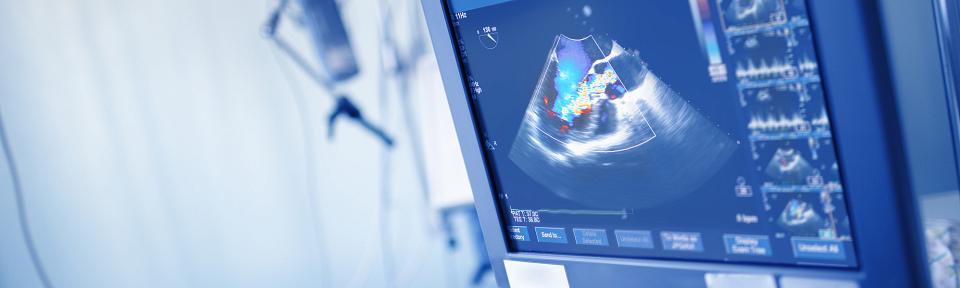
- Echocardiography
- Nuclear Cardiology
- Vascular Diagnostic Lab
- Vascular Ultrasound

Cardiac CTA (Computed Tomography Angiogram)
Coronary computed tomography angiograms (also called cardiac CTs) use contrast dye to create detailed, 3D images of the heart. These images show the heart moving as well as the large blood vessels inside the heart.
Doctors use cardiac CTAs to find blockages inside your coronary arteries. Your coronary arteries branch off of the aorta inside your heart and deliver oxygen-filled blood to your whole heart. It’s important for doctors to find any blockages inside your coronary arteries; if your coronary arteries are blocked, it will be more difficult for blood to flow to your heart. Over time, this could cause a heart attack or angina.
In the past, angiograms that looked at blood vessels inside the heart were fairly invasive. Our Cardiac Imaging Lab is proud to offer a newer alternative to the more invasive coronary angiogram. More doctors are recommending coronary CTA exams. This new technology can non-invasively detect soft plaque (fatty materials) inside artery walls that hasn’t hardened yet.
It’s important to find out if the arteries inside your heart have plaque inside them. Over time, this plaque may cause heart problems if you don’t make lifestyle changes or get medical treatment. In some cases, you may not need more invasive tests if you have a CT angiography.
Our Cardiovascular Center supports using CTA for many of our patients. But doctors won’t recommend this procedure for everyone because it won’t be useful in all cases. It’s very important that you speak with your doctor to find out whether this technology is right for you.
A dual source computer tomography scanner (CT) is a powerful, advanced test because it provides a clear, detailed picture of your heart. This CT scanner provides useful information to doctors. It allows our doctors to detect coronary artery disease even for patients who have no symptoms. Compared to older CT scanners, this leading-edge technology is safer because it's noninvasive and its speed minimizes exposure to radiation by almost 50 percent.
Cardiac MRI
U of U Health is the only medical center in the region that integrates the most advanced MRI imaging in treating heart disease. This allows our physicians to take a closer look at your heart and blood vessels with very low risk. Our advanced technology provides a better picture of complex heart abnormalities, giving a more in-depth diagnosis and treatment for each of our patients.
Nuclear Cardiology
Our team of specialists uses the latest technologies to screen patients who have a high chance of developing coronary artery disease but may not have any symptoms. Doctors can also use nuclear imaging to determine if patients with heart disease may benefit from interventional treatment.
Nuclear technology uses special radionuclide dyes to trace how the blood flows through the heart’s chambers, through the arteries that supply blood to heart, and through the heart’s muscles. This information allows our doctors to see how well a patient’s heart is pumping.
Patient Resources
Advances in Cardiology Imaging Mean Better Care
Cardiology imaging can give doctors crucial information to help them more confidently diagnose a patient and choose the best treatment. In this interview with Dr. Jeroen Bax, president of the European Society of Cardiology, learn more about the types of imaging available and ways doctors can best use them to detect and treat heart issues.