Anterior Skull Base Tumors
Traveling from Outside of Utah?
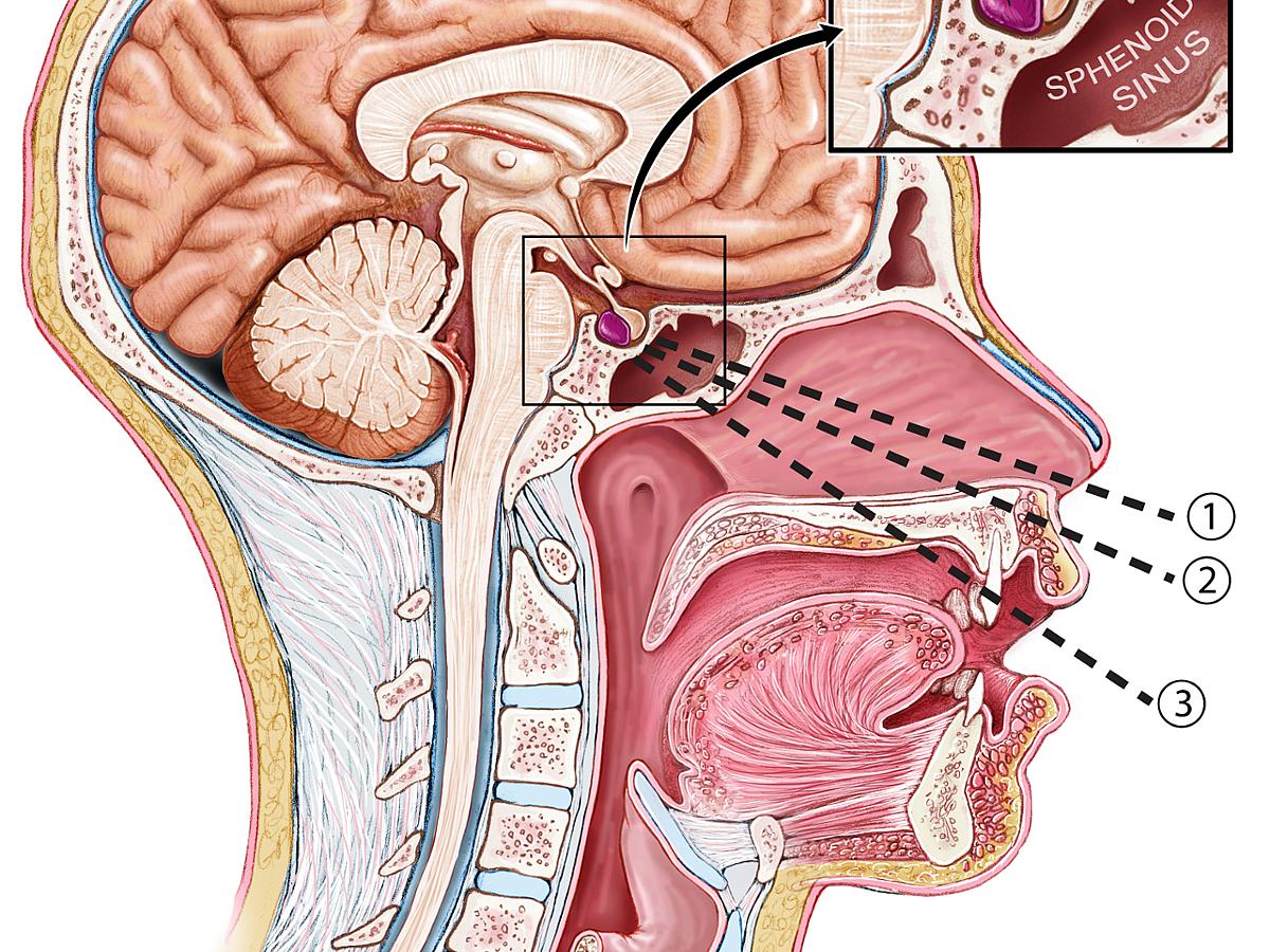
Anterior skull base tumors are found in the front of the skull base near where the eye sockets and sinuses are located. Some examples include:
- Meningiomas—A tumor found on the tissue covering the brain or spinal cord in the skull interior.
- Pituitary tumors—Tumors in the pituitary gland, which is located in the area behind the nose. They are also typically benign but can cause an increase or decrease in hormone levels.
- Esthesioneuroblastomas
- Sinonasal squamous cell carcinomas
- Sinonasal adenocarcinomas
- Sinonasal mucosal melanomas
- Sinonasal adenoid cystic carcinomas
- Sinonasal undifferentiated carcinomas
- Sinonasal neuroendocrine tumors
- Juvenile nasopharyngeal angiofibromas
- Inverted papillomas
Symptoms of Anterior Skull Base Tumors
Common symptoms include:
- vision and eye movement problems,
- weight loss,
- muscle weakness,
- reduced menstrual periods (pituitary tumors),
- erectile dysfunction (pituitary tumors),
- reduced sense of smell,
- nosebleeds,
- congestion (sometimes on one side),
- sinus infections, and/or
- problems breathing through your nose.
Find a Specialist
Anterior Skull Base Surgery
The location of the tumor, its size, and how it affects you, will help us determine if surgery is the best treatment choice. If so, there are two main approaches for anterior based tumors:
- minimally-invasive surgery involving an endoscope.
- traditional brain surgery, known as an open craniotomy, where we remove a piece of bone from the forehead or temple to access the tumor.
A team of doctors works together to remove the tumor and achieve the best outcome for you. Members of that team can include:
- a neurosurgeon,
- a rhinologist, and
- an anesthesiologist.
Endoscopic Endonasal Surgery
Skull base surgeons can remove many skull base tumors with the endoscopic endonasal approach. This is a minimally invasive technique involving an endoscope. Endoscopic end-nasal surgery allows our skull base surgeons to access tumors through the sinus and nasal cavities.
An endoscope is a medical device that transmits images via a long, thin tube and helps us examine the tumor. The endoscope is inserted into the nose. We then use microsurgical instruments beside the endoscope to cut out the tumor while carefully preserving the normal structures around it.
We may restore the area with healthy tissue from inside the nose or elsewhere in your body, such as your abdomen or leg, if needed.
During either or your procedures, we may need to temporarily install a spinal drain to divert any excess cerebrospinal fluid and help the area heal. We will remove the drain a few days after your surgery.
Craniotomy
In a craniotomy, we will access the brain by temporarily taking out a piece of skull. We will draw back the exposed brain during surgery to reach the tumor. We use this approach when we can't reach the tumor through endoscopic surgery.
During a craniotomy, we may view the tumor with the help of a large surgical microscope. Scans from prior imaging tests, which we use as a guide, help us know the exact location of your tumor.
At the completion of surgery, the small piece of skull is usually replaced.
Recovery After Skull Base Surgery
After surgery, patients may need to stay in the hospital for a period of four to seven days.
Medication can help relieve your pain and prevent brain swelling and seizure. Levels of exercise will vary, but many patients are encouraged to walk the day after surgery. We may ask you to limit heavy lifting for several weeks after surgery.
Our skull base surgeons will follow up with you to monitor changes in symptoms. More imaging tests will be needed.
Most patients are able to return to work and normal daily activities after recovery.