As the Project Administrator of Ophthalmic Imaging at the Moran Eye Center, Jim Gilman has established a reputation for both technical mastery and artistic skill, winning more than 40 awards for his work since 1982.
Gilman’s expertise was once again acknowledged at the November 16-19 2013 Ophthalmic Photographers’ Society Annual Scientific Exhibit at the American Academy of Ophthalmology (AAO) in New Orleans. Judges selected three of his images among a field of almost 600 entries from around the globe."You can tell I’ve been doing this for a while," says Gilman. Over time you can recognize when an image is special. Whether the pathology is unique or the view in the eye is spectacularly clear. When the medical scientific information combines with an artistic aesthetic, these images take on a higher value as powerful windows to the human body to be shared and admired.
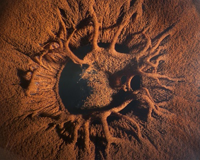
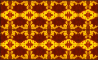
First Place in the "Cross Category" section, is his print of a ciliary body tumor using a gonio lens and a slit lamp photo combined. Second Place in the "Slit Lamp Photography" Category, the papillary membrane that appears almost like a lunar landscape. Second Place in "The Eye as Art", is Gilman's mosaic, created on a fundus photograph of serpiginous and altered to resemble a Persian carpet. (A fundus photo is a color picture of the retina, taken with cameras specially designed to look optically through a dilated pupil and flatten a curved surface in the back of the eye.)
The medical imaging specialists at the Moran ophthalmic imaging and videography department use technology, photography, art, medicine, and science to create pictures and videos of the inside and outside of the eyeball every day. The images are stored in an electronic database where they can be accessed and studied by the physicians. These images can be viewed from different angles, sent to other doctors or departments, or safely saved and stored for future use, all of which aid in treating everything from low vision to blinding eye diseases.
Some of the tests performed at Moran include spectral domain optical coherence tomography (OCT, which is a scanning laser procedure), auto fluorescence, blue reflectance, infrared, color fundus photography, ICG and fluorescein angiography, slitlamp biomicrography, goniography and external photography.