While ophthalmic imaging technology becomes more and more sophisticated each year, photos of the eye seem to take on other worldly specters: a lunar eclipse, a Martian crater, or the Galilean Moons of Jupiter. And while these images help doctors and researchers make discoveries, diagnoses, and treatments, they also reveal dramatic, organic patterns, as cosmic looking as a solar flare or as abstract as a cat's-eye marble.
The Ophthalmic Photographers Society recently awarded top honors to five Moran Eye Center photos out of 900 submissions during its Scientific Session held at the American Academy of Ophthalmology (AAO) annual meeting. Accolades go to Moran's Jim Gilman, Melissa Chandler, Becky Weeks, and Danielle Princiotta.
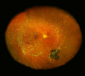
"Asteroid Hyalosis" Jim Gilman, CRA, FOPS, First Place, Ultra-widefield Imaging.
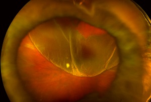
"Choroidal Rupture" Melissa Chandler, COA, CRA, OCT-C, Second Place, Monochromatic Photography.
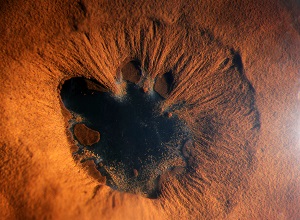
"Giant Retinal Tear" Jim Gilman, CRA, FOPS, Third Place, Ultra-widefield Imaging.
"Posterior Synechiae" Becky Weeks, COA, CRA, Honorable Mention, Slit Lamp Photography.
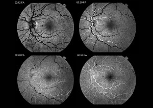
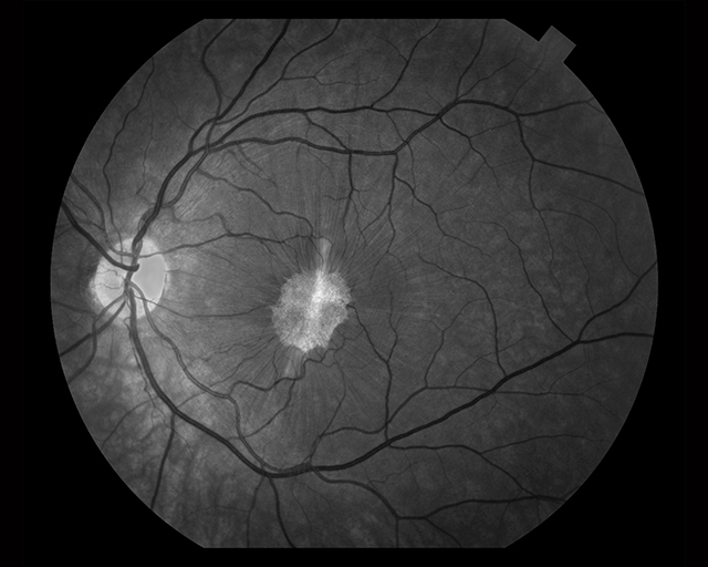
"Central Retinal Vein Occlusion" Danielle Princiotta, COA, Honorable Mention, Fluorescein Angiography.
The awards were in addition to five Moran physicians who received top honors at AAO.