Long before retinal diseases begin to rob a person’s eyesight—long before symptoms develop or a thorough dilated eye exam can discover it—retinal cells begin a quiet, unnoticed struggle to survive.
It’s only after a substantial number of cells have died that diseases including glaucoma, age-related macular degeneration, and rarer inherited conditions are discovered and treated.
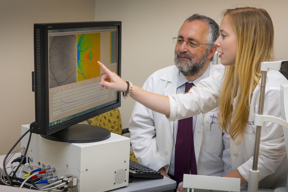
But a new imaging tool, brought to the John A. Moran Eye Center researchers by visiting international physician Lydia Sauer, MD, suggests a potential shift in the paradigm. An extremely sensitive, non-invasive camera—the first of its kind in the U.S.—has allowed retinal specialist Paul S. Bernstein, MD, PhD, to identify subtle changes in retinal cells that point to disease earlier than ever before.
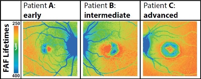
Using the fluorescence lifetime imaging ophthalmoscopy (FLIO) camera, Bernstein, Sauer, and Moran resident Rebekah Gensure, MD, PhD, identified macular telangiectasia type 2 (MacTel)—a notoriously difficult to diagnose hereditary disease that causes central vision loss. FLIO not only showed a metabolic cell ‘signature’ for early stage MacTel, but evidence of a difference in the metabolic state of retinal cells in people who are likely to have a carrier gene but no symptoms of the disease.
"We were able to image more than 20 people with MacTel for the study, and that’s a large number for a rare disease," said Bernstein. "FLIO has a great future as an imaging technique, and MacTel appears to be an ideal condition to demonstrate its potential."
Since the actual gene responsible for MacTel hasn’t yet been found, genetic testing isn’t available. But FLIO, in the hands of experts, could ferret out a person’s risk for developing it.
FLIO might also someday be used, for example, to identify the onset of a host of other inherited eye diseases.
WHAT IS FLIO?
The FLIO camera records the mean lifetimes of fluorophores, a fluorescent chemical compound found in the retina that re-emits light when it is stimulated by a laser. These measurements show the health of retinal cells.