
Thomas D. "Tom" Dee II first found himself in the office of Randall J Olson, MD, in the 1990s.
Olson told Dee he showed early signs of age-related macular degeneration (AMD), and there was no cure for the disease, which destroys central vision.
"Tom experienced firsthand how bad the scourge of macular degeneration can be," Olson said. "He was also one of the first to want to change that reality."
Dee’s friendship with Olson, and his experience dealing with AMD, led him to become one of the John A. Moran Eye Center's biggest advocates for research fighting the eye disease. He supported Moran’s AMD research until his passing in 2009. His sons, Tim and David Dee, and Tim’s sons Matt and Nate now lead the family’s philanthropic efforts through their foundation, the Lawrence T. & Janet T. Dee Foundation, named after Tom’s parents. They’ve remained involved with Moran, supporting AMD research.
"For us, this idea of being a part of health care in the community is just a part of family culture," David Dee said. "It brought a lot of satisfaction to our grandfather and our father."
The Dees have been behind several gifts crucial to advancing the fight against AMD, including funding the recruitment of Gregory S. Hageman, PhD, whose lab is devoted to finding new therapies for the disease.
The Dee Family's Legacy of Giving
The Dee family has been investing in the Utah health care community for nearly a century. Annie Taylor Dee of Ogden dedicated her life to the pursuit of better medical services after losing her husband and oldest son to preventable causes.
Her vision for improving Utah’s health services has been transformational: McKay-Dee Hospital in Ogden exists due in part to Annie’s efforts and generosity, and the nursing school at Weber State University bears her name.
Annie’s great-grandchildren continue to honor her legacy.
"I would say our values, vision, and mission are evolving, and we’re bringing on the next generation," David said. "But we always try to honor what our dad and our grandparents cared about."
Next-Generation Projects to Fight Blinding Eye Diseases
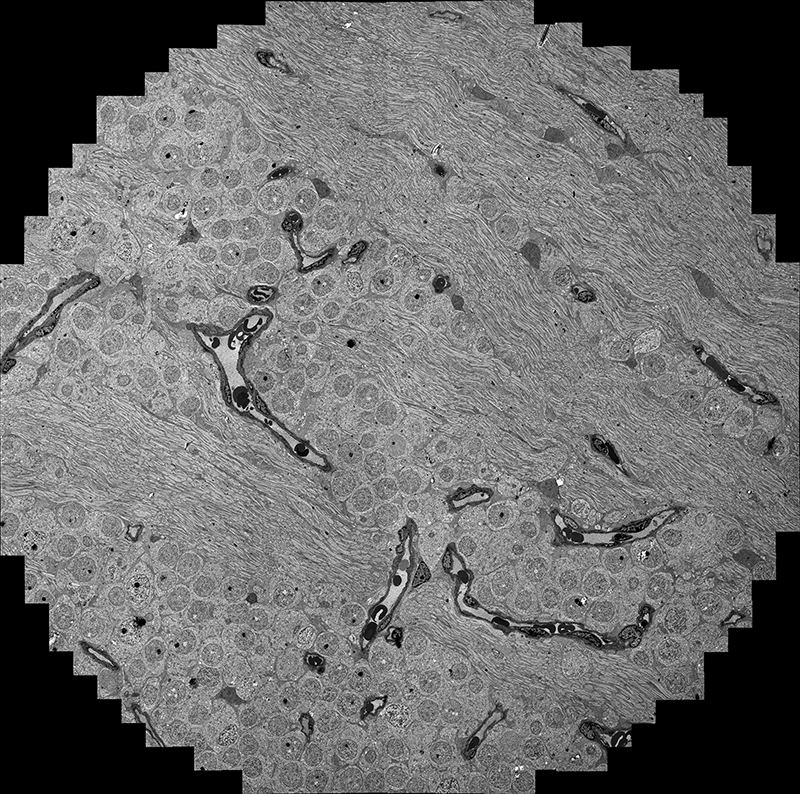
It was the next generation, Matt and Nate, who suggested the Dee Foundation fund research equipment at Moran, including a transmission electron microscope. This microscope enables the visualization of the connections between neurons in the eye for the study of neural connections, called "connectomics." These connections between neurons are smaller than the wavelength of light and cannot be observed with regular light microscopes.
Bryan W. Jones, PhD, director of Moran’s Marclab for Connectomics, said the new microscope has transformed his research mapping the circuitry of the retina, including a new project to map corruption in circuitry that occurs in AMD and other blinding diseases.
"The addition of the second, more efficient microscope has effectively more than doubled our output," he said.
The Dee Foundation has had a profound impact at Moran, the University of Utah, and the greater Utah community.
Researcher Spotlight: Bryan W. Jones
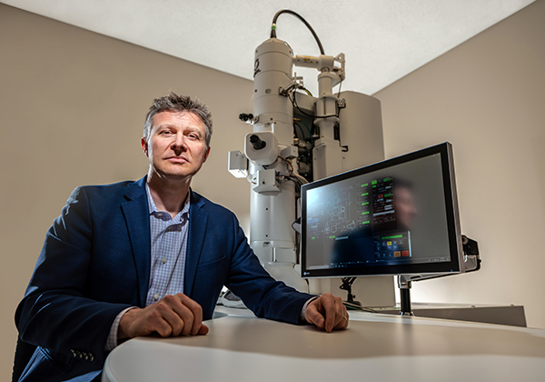
Bryan W. Jones, PhD, heads the Marclab for Connectomics at the Moran Eye Center. Connectomics is the study of neural connections—specifically, in Jones’s case, in the retina. The Marclab, then under the direction of Robert E. Marc, PhD, was the first in the world to build a map of the circuitry of the retina, and that original map is still being explored and continues to provide vital research data.
Jones’s lab has two transmission electron microscopes, working nonstop nearly every day of the year, producing an unprecedented amount of data to understand retinal circuitry. His lab is the only National Institutes of Health-funded connectomics research group, competing with much larger privately funded groups yet managing to produce more publications on the retinal connectome than any other institution.
Recently, Jones received a multiyear National Eye Institute (NEI) grant totaling more than $3 million to research the next step in connectomics—the "patho-connectome," or the study of how diseases progress through the retinal circuitry.
With the new electron microscope and the backing from the NEI, Jones is looking to unravel the mysteries of eye diseases and provide data that could be an invaluable asset for the vision science community.
"The support of the Dees has kept Moran competitive with the largest institutions in the world engaged in connectomics research."
Bryan W. Jones, PhD, director of Moran’s Marclab for Connectomics