Exploring New Frontiers of Retinal Imaging
In the future, a two-minute retina scan could let doctors spot eye diseases—or even the risk to develop them—long before any symptoms appear.
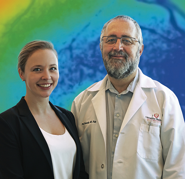
"With more lead time, therapies and interventions have a much better chance to stop or slow disease," said Paul S. Bernstein, MD, PhD, with the John A. Moran Eye Center. "It’s what every doctor hopes for."
This future is becoming more likely as Bernstein and research associate Lydia Sauer, MD, conduct studies using a remarkable new non-invasive retinal camera. From age-related macular degeneration (AMD) to diabetic retinopathy, drug toxicity, and inherited retinal conditions, Bernstein’s lab has made groundbreaking discoveries over the past two years using the only Heidelberg Engineering Spectralis-based fluorescence lifetime imaging ophthalmoscope (FLIO) in the U.S.
Using FLIO, Bernstein and Sauer are mapping subtle retinal changes that could one day also indicate glaucoma, uveitis, Alzheimer’s disease, and many other conditions.
How FLIO Works
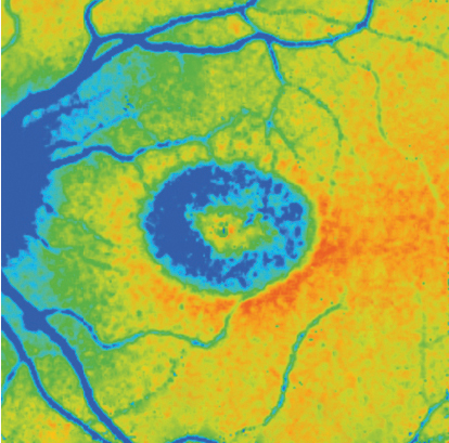
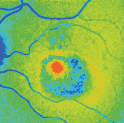
FLIO allows doctors to examine the health of the retina by measuring how long molecules known as fluorophores emit light, or glow, when stimulated with a laser. This is known as the autofluorescence lifetime of the fluorophore.
Areas with the shortest autofluorescence lifetimes are color- coded red; the ones with the longest are blue. Healthy eyes show typical areas of red and blue, but these patterns change in different eye diseases, creating unique signatures that indicate diseases, disease progression, and even risk of developing certain diseases.
Bernstein and Sauer first used FLIO to study macular telangiectasia type 2 (MacTel), a rare inherited eye disease that causes a gradual loss of central vision. Working with a large study group of families with the disease as part of an international research project, the Moran team not only identified a FLIO signature for early-stage MacTel but also discovered changes in people with a genetic risk of developing the disease. Additionally, FLIO played a role in helping Bernstein and fellow MacTel Project scientists and clinicians around the world identify the first genetic cause of the eye disease in 2019.
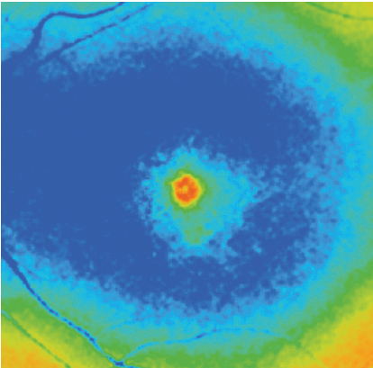
One of the biggest potential clinical uses of FLIO could be the early diagnosis of AMD, a leading cause of central vision loss among adults age 55 and older in the U.S. Bernstein’s lab was the first to describe a specific signature ring pattern in FLIO images of eyes with AMD.
"We see this ring pattern in all of the AMD patients we’ve tested," Sauer said. "And it appears to be getting stronger as the disease progresses."
FLIO has also shown a trace of the ring pattern in a few patients who seem to be healthy using other imaging techniques. "We believe we are seeing patterns of AMD even before a person is showing any symptoms," Sauer said.
New Screening Potential
Bernstein and his lab recently published promising results of using FLIO to screen for retinal toxicity caused by hydroxychloroquine (brand name Plaquenil). Doctors prescribe the drug to prevent malaria and treat dermatologic and rheumatologic inflammatory conditions such as rheumatoid arthritis and lupus.
"The problem is that if you’re on the drug for more than five years, about 10-20 percent of people develop toxicity that can actually blind them," Bernstein explained.
Working with the University of Utah’s Department of Neurology, Bernstein is also exploring FLIO’s potential to detect Alzheimer’s disease. The theory is that the same kind of protein deposits seen in brain scans of Alzheimer’s patients may be detectable in the eye. If that’s the case, FLIO could potentially replace more costly brain imaging methods now used for Alzheimer’s.
FLIO Research Expands Internationally
FLIO, developed in Germany, has been available only in a few centers in Germany, Switzerland, and at Moran. Heidelberg will soon release an upgraded version of FLIO to Moran and an expanded list of about a dozen centers in the U.S. and worldwide. Moran is poised to help lead a research consortium coming with the expanded release.
"The sites will share data, work together, see how different diseases manifest in different parts of the world," said Bernstein, "as well as use artificial intelligence technology to improve data analysis."
All the researchers who have worked with FLIO so far believe it has incredible potential, Sauer said. "With this new technique, we may understand eye diseases much better in the future."