The National Science Foundation (NSF) has awarded John A. Moran Eye Center scientist Bryan W. Jones, PhD, funding as part of an international group of neuroscientists that will explore the connections between neurons, known as synapses.
Jones, director of the Marclab for Connectomics at the Moran Eye Center, studies neural connections in the retina. The lab was the first in the world to build a synaptic level map of the circuitry of the retina and is known for its ongoing discoveries in how retinas change in retinal degenerative disease.
Jones now joins one of four interdisciplinary teams receiving more than $50 million over five years as part of the NSF’s Next Generation Networks for Neuroscience program, also known as NeuroNex. A total of 70 researchers, representing four countries, will investigate aspects of how brains work and interact with the environment around them.
Understanding the Brain to Better Treat Eye Diseases
Collaborating with the laboratory of University of Utah Department of Biology Distinguished Professor Erik Jorgensen, PhD, and a team based at the University of Texas, Austin, Jones will work on a project examining the relationship between the weight, or strength, of synapses and their structural components. In the retina, synapses allow for the communication between neurons involved in the detection of light and the processing of information associated with vision, such as contrast, color, and movement.
"Understanding how the synapses change in retinal disease is crucial to understanding how to design therapeutic interventions," Jones explained. "For a long time, it was presumed that the communication between neurons at synapses was an all or nothing event. But now we know that synaptic communication is much more nuanced."
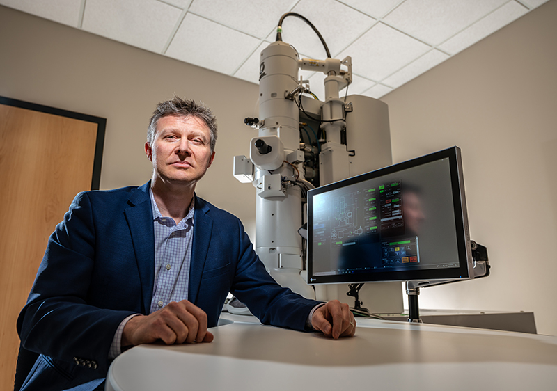
Jones will study how synapses are altered in retinal degenerative disease using electron microscopes to reconstruct and analyze synapses.
"Our second and newest electron microscope, recently gifted by the Lawrence T. & Janet T. Dee Foundation, will be crucial to this effort," said Jones. "It provides a sensitive camera and another transmission electron microscope that we will need to visualize synapses at multiple angles and to perform the work required for this grant."