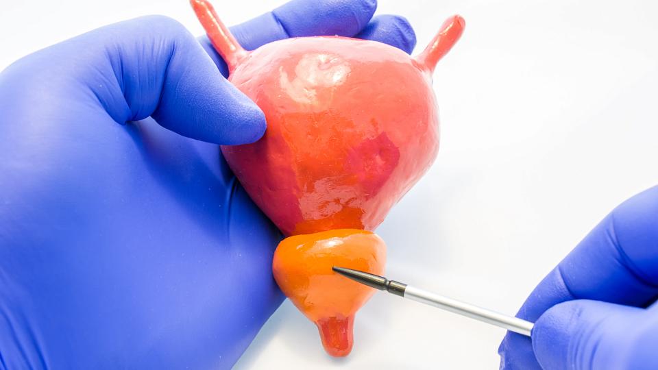
What Are Ureteral Obstructions and Strictures?
A ureteral obstruction is a blockage in your ureters. Your ureters are the tubes that carry urine from your kidneys to your bladder. Many conditions, including kidney stones, cancer, and ureteral strictures can cause an obstruction.
Ureteral strictures are when part of your ureter becomes scarred and narrowed. Ureteral blockages and strictures are often treatable. But, without treatment, they can cause pain, kidney stones, urinary tract infections, kidney damage, and even kidney failure.
Ureter vs. Urethra
Both your ureters and urethra play important roles in expelling urine from your body. The ureters take urine to the bladder. The urethra is the small tube that carries urine from your bladder outside your body. Scarring and narrowing can also affect your urethra. This is known as a urethral stricture.
Find a Urologist
Ureteral Stricture Symptoms
Some ureteral strictures cause only mild symptoms or no symptoms at all. The signs of a ureteral stricture depend on multiple factors, including how severe the blockage is and whether it affects both ureters.
Patients who do have symptoms of an obstruction may notice a few things:
-
Frequent urinary tract infections (UTIs)
-
Pain between your upper belly and back (flank pain)
-
Pain in your kidneys, which feels like back pain
In severe cases, urine backup may cause your kidneys to swell. This is known as hydronephrosis. Blockages that don’t get treated can damage your kidney function.
Causes of Ureteral Obstructions
There are several causes of ureteral obstructions:
-
Abdominal cancer that pushes on your ureters
-
Developmental problems that are present at birth (congenital)
-
Inflammation in your abdomen and the tissues around your ureter (retroperitoneal fibrosis)
-
Ureteral strictures
Ureteral Strictures After Surgery
One of the most common causes of ureteral obstruction is scarring (strictures). This scarring may develop after surgery to an area close to the ureters:
-
Gynecologic surgery, such as removal of the ovaries or uterus (hysterectomy)
-
Urologic surgery for kidney stones
-
Vascular surgery on your abdominal arteries
Uretero-Pelvic Junction Obstruction
Your kidney and ureters join in your upper abdomen. This area has two parts:
-
Renal pelvis: The funnel that collects urine from your kidney
-
Uretero-pelvic junction: The area where the kidney and ureter connect
Sometimes, the way your kidney develops before birth blocks the uretero-pelvic junction. Other times, these obstructions arise later in life.
These blockages may occur for several reasons:
-
Blood vessels that cause a kink in the uretero-pelvic conjunction, preventing urine from draining well
-
Ureteral strictures
-
Ureters that don’t function as they should
Treatments for Ureteral Obstructions and Strictures
Treatment for a ureteral obstruction varies based on the underlying cause. Sometimes treatment starts with procedures to help urine drain from your body:
-
Nephrostomy tube: We make an opening in the skin near your kidney and insert a tube that allows urine to drain. Our interventional radiologists perform this procedure.
-
Ureteral stenting: Your surgeon places a hollow tube inside your ureter to open it. This may be temporary treatment before surgery.
Your urologist may be able to treat the underlying cause of the blockage after addressing the urine drainage. They may use one or several treatments:
-
Balloon dilation, a procedure that uses a catheter (drainage tube) and balloon to widen a ureteral stricture
-
Surgery to remove the blockage and repair your ureter
Why Choose University of Utah Health?
University of Utah Health urologists are experts at treating ureteral obstructions and strictures. We combine clinical expertise with the latest research findings and state-of-the-art equipment. Our surgical specialists provide patients with the highest level of care available in the Mountain West.
Make an Appointment
Call our clinic at 801-213-2700 to make an appointment. You don't usually need a referral to see our urologists. If your provider would like to make a referral, they can fill out our referral form.
Hear From Our Patients
Pat O’Neal underwent radiation therapy when his prostate cancer spread. However, the radiation damaged his ureters (tubes that move urine from the kidneys to the bladder). He soon experienced urinary issues. After trying other treatments that negatively impacted his quality of life, he came to U of U Health for reconstructive ureteral surgery.