Diagnosing Epileptic Seizures
For about two-thirds of patients with epilepsy, seizures can be effectively treated or managed with the use of anti-seizure medication. However, one-third of patients continue to experience seizures even on medication. If your medication is not helping you manage your seizures, your neurologist may recommend intracranial monitoring -- a surgical procedure to learn more about the location of your seizures.
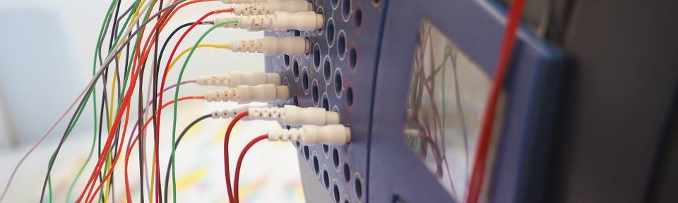
Intracranial monitoring is an invasive surgery where neurosurgeons place electrodes inside your brain to monitor brain activity over several days or weeks. Your neurologist will opt for intracranial monitoring if the initial test results didn't provide enough information.
Goals of Intracranial Monitoring
While it is an invasive procedure, intracranial monitoring will provide doctors with valuable information in the following ways.
- Finds the exact precise location of seizures.
- Finds tissues that could be removed to prevent seizures.
- Maps out the patient’s brain to help protect critical functions, like speech and movement.
Once intracranial monitoring is complete, your neurosurgeon will consult with you and let you know whether surgery is safe and is the best option to treat your seizures.
Preparing for Surgery
Before surgery, your doctors will use non-invasive test results from MRI (magnetic resonance imaging) scans or neuropsychological evaluations to identify the area of your brain where they believe the seizures are occurring. Your pre-surgery meeting may include some or all the following members of a care team:
- epilepsy specialist,
- neurosurgeon,
- neuropsychologist,
- neuroradiologist, and
- other specialists involved in your care.
Your epilepsy treatment team will create a map of where to place the intracranial monitoring electrodes. Their goal is to gather the best data and find the location where your seizures are occurring.
Before the surgery, you should discuss all medications you are currently taking with your doctor including:
- prescriptions,
- over-the-counter drugs,
- vitamins,
- supplements, and
- herbal products.
Your neurologist may recommend that you stop or make changes to some or all your medications before intracranial monitoring. Make sure to tell your doctor about any blood thinners you are taking such as aspirin. This helps reduce the risk of bleeding. Do not stop or change any medications without consulting your physician.
Find a Provider
Surgery: Electrode Placement
There are two types of intracranial surgery: subdural (space between the brain cover and the brain itself) electrodes and depth electrodes (stereo-EEG). The one you get depends on where neurologists believe the seizures start inside your brain. You will be put to sleep under general anesthesia during these surgeries to place the electrodes, which typically takes a few hours.
Subdural Electrodes (Strips or Grids)
If your neurologist believes the seizure location is somewhere on or near the brain's surface, they will use subdural electrodes. These are placed under the dura, or the cover of the brain. Several electrodes are either arranged in a strip (a long rectangle) or a grid pattern (a square).
Subdural electrodes need to be placed using a more invasive procedure called a craniotomy. During this surgery, your neurosurgeon will temporarily remove part of the skull to place the strips or grids on the brain. The bone is then replaced and skin stitched closed. The electrodes are connected with wires to a device that will remain with you until the intracranial EEG monitoring is complete.
After your electrodes are placed, you are taken into a recovery unit (usually for one night), and finally to an epilepsy monitoring unit for the intracranial EEG.
Depth Electrodes (Stereo-EEG)
If your neurologist believes the seizures are located deep inside your brain, they will implant depth electrodes (thin, needle-like wires) into your brain. These electrodes send information from multiple points along the electrode during monitoring.
Your neurosurgeon will secure your head with a metal frame. The electrode entry points are then targeted using a small robot, or manually entered into the frame. Small holes are drilled in your skull at the places they identified, and put the electrodes through the holes. Small wires connect the electrodes to a portable pack that will remain with you during the monitoring procedure.
Surgery Risks
Both types of electrodes require a surgical procedure and come with some risks. The most common ones are:
- bleeding,
- brain swelling (edema),
- infection,
- neurological impairments (these are rare), and
- post-operative headaches or pain.
Stereo-EEGs require smaller incisions in a less invasive procedure. They often result in fewer complications, less pain, and lower risk of infection or complications after the operation. Your neurosurgeon will likely prescribe antibiotics after surgery to reduce the risk of infection.
Intracranial EEG Monitoring
Intracranial EEG monitoring could last anywhere from a few days to three weeks. The electrodes will remain in place this entire time. Your electrodes are attached to wires and connected to a portable pack that allows you to move around the room during the monitoring procedure. You should bring activities to keep you busy and avoid boredom during the test because it will last for at least several days.
During monitoring, your neurologist and neurosurgeon will send tiny electrical currents along the electrodes and watch for changes in:
- vision,
- movement,
- language, and
- other critical functions.
This information creates a “map” of the brain to determine whether surgery is safe. It also helps surgeons avoid these areas during the procedure.
A neurologist will watch to see what happens before, during, and after a seizure to find the precise seizure location. When your team gathers enough information, the electrodes are removed. Depth electrodes can often be removed in a simple exam room procedure while you are awake or under light sedation. (where you are put to sleep).
Recovery
After your monitoring and surgeries are complete, you may either leave the same day, or stay for additional nights. Once you are discharged, the staff will schedule a follow-up appointment for you to see the neurosurgeon or neurologist.
Incisions should be kept dry for 72 hours after your surgery. Incisions usually heal in about 10 to 14 days. Sutures (stitches), if you have any, should come out at this same time.
Make an Appointment with Our Neurologists
You will need a referral from your primary care physician or another provider to be seen in our Epilepsy Clinic. Call 801-585-7575 to schedule an initial appointment after you are referred.
Depending on your treatment plan, you will see an epileptologist for evaluation and work with the surgical coordinators to schedule any necessary tests or procedures for diagnosis and to determine if surgery is the best option. If surgery is part of your treatment plan, our team will help coordinate your care and schedule an appointment for you with a neurosurgeon.


